


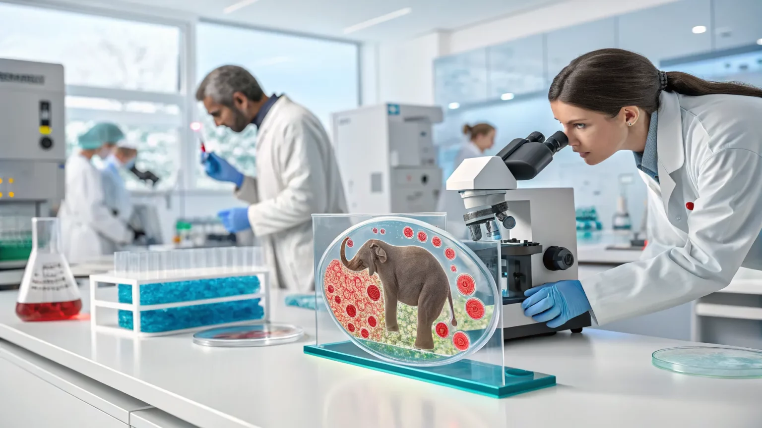
In a first for bioengineering, a research team has 3D-printed microscopic shapes inside living cells, including a tiny elephant and scannable barcode tags. The work, described this week, signals a new phase for building tools within life’s basic units. It could change how scientists track cells, deliver drugs, and study disease.
The experiments took place in lab settings with cultured cells. The printed objects were created on the scale of micrometers, small enough to sit inside a cell without piercing its membrane. The group’s goal was to show that complex, predesigned objects can be formed in place, while the cell remains alive.
“For the first time, researchers have 3D-printed tiny objects directly inside of living cells, including an elephant and barcode tags.”
What Happened
The team produced two types of objects that show both artistry and utility. The elephant demonstrates geometric control at a minute scale. The barcodes hint at practical applications: objects that can be read later to identify, sort, or track cells.
Earlier approaches usually built structures outside the cell, then inserted them by microinjection or uptake. Those steps can stress cells and limit shape complexity. Printing inside cells aims to skip these transfer steps and shape materials precisely where they are needed.
How The Printing Likely Works
While full technical details were not released, the process likely uses focused light to harden a liquid material inside the cell. In similar micro-printing methods, a laser activates a photosensitive gel at exact points, forming a solid line by line. The printing path creates a 3D object without moving the cell.
Keeping cells alive through this process is the core challenge. It demands careful control of light intensity, exposure time, and chemical ingredients. Early results suggest that at least some cells tolerate the procedure, though long-term effects need testing.
Why It Matters
Printing inside living cells opens new paths for research and medicine:
- Custom labels: Barcodes can tag single cells so their history can be read later.
- On-demand sensors: Tiny structures could report on pH, ions, or mechanical forces inside cells.
- Therapy platforms: Internal scaffolds may carry drugs or guide how a cell grows and divides.
Cell tracking is a long-standing hurdle in fields like cancer, immunology, and stem-cell science. Tools that stay inside the cell and endure many divisions could help map cell fate with higher accuracy. If barcodes persist through cell cycles, labs could watch how a single cell gives rise to many, with less guesswork.


Context And Precedents
Micro 3D printing has matured over the past decade, allowing intricate features smaller than a human hair. Two-photon lithography and similar techniques have built tiny scaffolds, valves, and optics. But most constructs sit on chips or inside droplets, not within living cells.
Researchers have also used DNA origami, nanoparticles, and protein tags to shape or label the cell interior. Each method trades off control, durability, and toxicity. Intracellular printing seeks more complex shapes with spatial precision and fewer insertion steps.
Safety, Ethics, And Limits
Key questions remain. How many cells survive the process? Do printed objects disturb normal functions, like gene expression or metabolism? Can the immune system react if these cells are used in animals or patients?
Ethical discussions will grow as the method advances. Permanent tags inside cells raise privacy issues if used in human samples tied to identity. Oversight boards will expect clear rules on consent, data handling, and limits on clinical use.
There are technical limits too. Large prints could crowd the cytoplasm or block transport. Materials must be stable yet safe to break down if needed. Readout tools must detect barcodes reliably across many cell types.
What To Watch Next
Near-term studies will focus on cell health and print fidelity over time. Teams will test different materials, shapes, and cell types, from immune cells to neurons. If barcodes can be read with standard microscopes or gentle imaging, the method may spread quickly across labs.
Regulators will look for dose limits on light and chemicals, plus evidence that edits are reversible or safe in animals. Partnerships between bioengineers and clinicians could push pilot studies in tissue models and organoids.
This debut shows that complex, readable structures can be formed inside living cells. The next phase is proving they help answer hard questions in biology and medicine, without harming the cells they are meant to guide.