





Ada Yonath spent years pursuing a scientific goal that many colleagues dismissed as a dead end. Her work mapping the ribosome later helped researchers design antibiotics and earned global recognition.
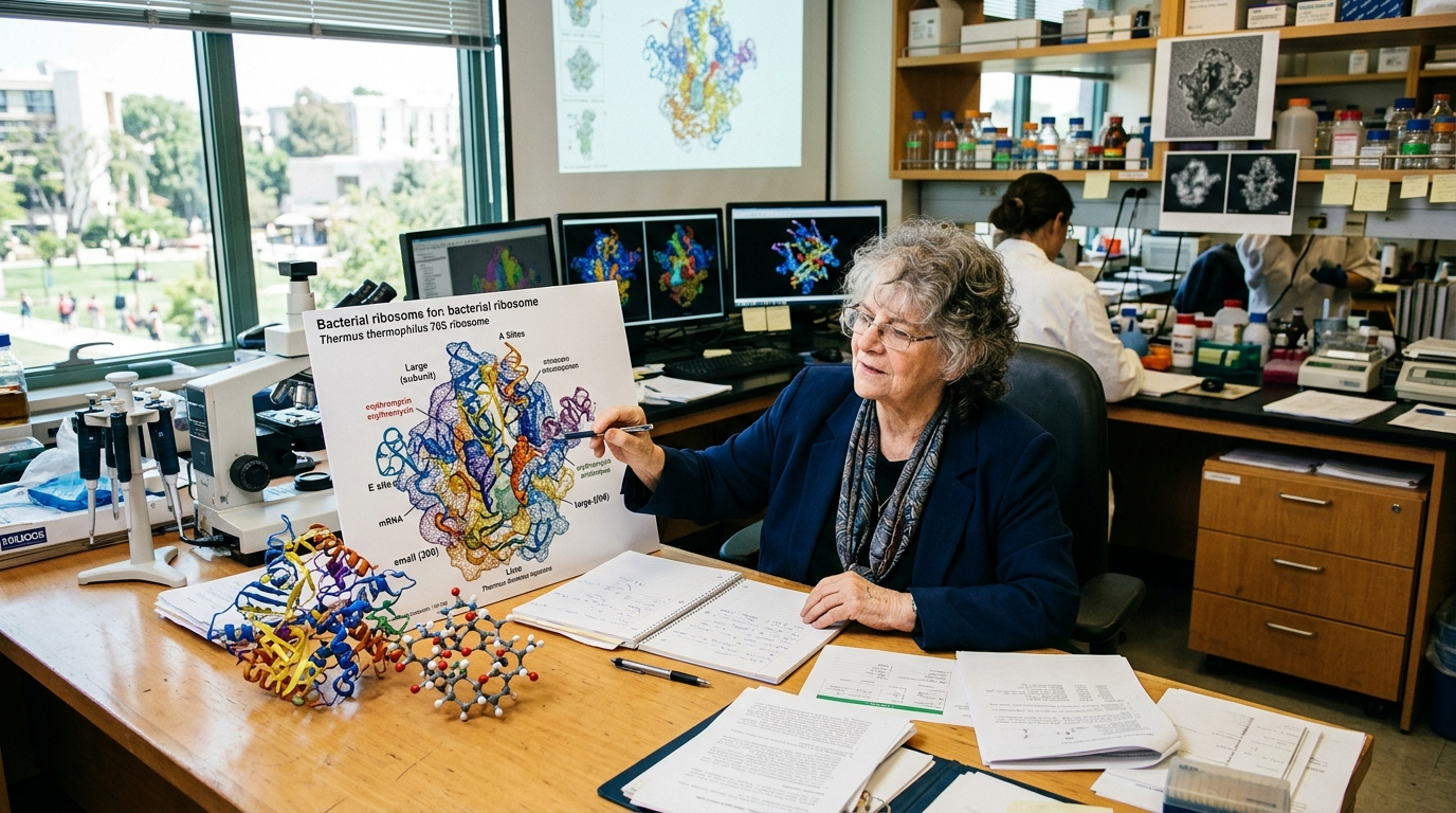
The Israeli crystallographer sought to reveal the ribosome’s three-dimensional structure at atomic scale. Ribosomes are tiny molecular machines that read genetic instructions and build proteins. Because bacterial ribosomes are targets for many antibiotics, seeing their structure gave scientists a clearer guide for drug research.
A Difficult Scientific Target
Yonath began studying ribosomes in the late 1970s. At the time, researchers struggled to produce stable ribosome crystals needed for X-ray crystallography. The structures were large, flexible and easily damaged.
Many scientists doubted that the method would produce useful results. Her effort “faced years of derision,” with critics viewing the project as unlikely to succeed.
Yonath persisted despite that skepticism. She developed techniques to crystallize ribosomes and protect samples from damage during experiments. Her team’s early results helped establish that detailed structural images were possible.
Her mapping of the ribosome, which led to new designs for antibiotics, faced years of derision, as many scientists saw it as a dead-end effort.
The resistance reflected the scale of the challenge. A ribosome contains RNA and proteins arranged in two main subunits. Mapping those parts required extensive testing and increasingly precise measurements.
Maps Reveal How Antibiotics Work
Yonath’s research helped show where antibiotics attach to bacterial ribosomes. Many such drugs block protein production, which can stop bacteria from growing or kill them.
Atomic-scale maps allowed researchers to examine several key questions:



- Where specific antibiotics bind inside a bacterial ribosome.
- How that binding disrupts protein production.
- Why genetic changes can make bacteria resistant.
- How future drugs might target bacteria more precisely.
These findings did not produce a simple cure for antibiotic resistance. Drug development still requires laboratory studies, safety testing and clinical trials. Yet structural information gave medicinal chemists a stronger basis for modifying compounds and comparing possible targets.
The work also helped explain why some antibiotics harm bacteria while largely sparing human cells. Bacterial and human ribosomes perform similar jobs, but their structures differ. Those differences can guide the search for treatments with fewer harmful effects.
Recognition Followed Years of Doubt
In 2009, Yonath shared the Nobel Prize in Chemistry with Venkatraman Ramakrishnan and Thomas Steitz. The award honored their studies of the ribosome’s structure and function.
Yonath became the first Israeli woman to receive a Nobel Prize. She was also the first woman in 45 years to win the chemistry prize, placing her achievement within a wider debate about women’s recognition in science.
The shared award reflected the role of several research groups. Ramakrishnan and Steitz led major structural studies that added detail to the scientific picture. Together, the findings changed how researchers understood one of the cell’s most basic processes.
A Continuing Fight Against Resistance
The research remains relevant as bacteria evolve resistance to existing medicines. Misuse and overuse of antibiotics can speed that process, leaving doctors with fewer treatment choices.
Ribosome maps offer one route for studying resistance and designing drugs, but they are not a stand-alone answer. Public health measures, responsible prescribing and investment in new treatments remain necessary.
Yonath’s experience also shows how uncertain research can yield practical gains. A project once mocked as impractical produced knowledge that influenced antibiotic design and basic biology. As resistant infections spread, the structures she helped reveal will remain an important scientific guide.